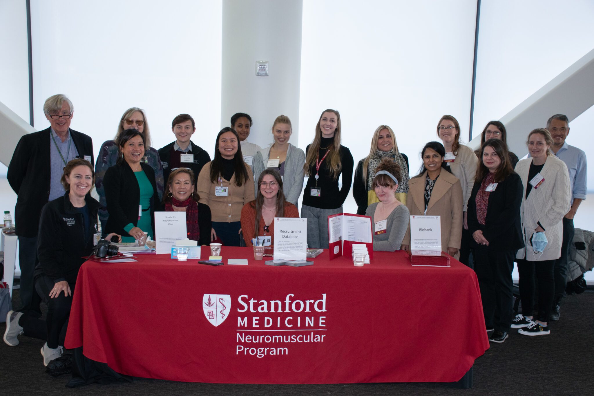
The SMA Researcher Meeting is the largest research meeting in the world specifically focused on SMA. This year we had a record setting 350 attendees. The goal of the meeting is to create open communication of early, unpublished scientific data, accelerating the pace of research. The meeting also furthers research by building productive collaborations—including cross-disciplinary dialogue, partnerships, integration of new researchers and drug companies, and educational opportunities for junior researchers.
We will be posting a series of summaries from our 2016 researcher meeting, highlighting some of the most interesting new developments and discoveries presented there. This update covers a session on SMN protein. Individuals with SMA do not correctly produce survival motor neuron (SMN) protein at high enough levels, due to a genetic mutation in the SMN1 gene. All patients with SMA have at least one copy of a low-functioning “backup gene” called SMN2. SMN2 cannot prevent SMA because it is misspliced, meaning it primarily produces a shortened, less functional SMN protein. However, when SMN2 is correctly spliced, it is able to produce some fully functional SMN protein. Understanding how to promote the correct splicing of SMN2 is valuable for therapeutic development, as is understanding when, where, and why SMN protein expression is needed in the body.
This session was moderated by Cure SMA Scientific Advisory Board Member Rashmi Kothary, PhD.
There were six talks in this session. Dawn Chandler, PhD (Ohio State University), presented on how stress due to lack of proper oxygen supply to tissues increases during disease in a mouse model of SMA. This lack of oxygen correlates with further worsening of SMN2 missplicing, generating less functional SMN protein. Of interest, another stress inducer, heat shock, actually improved SMN2 splicing and SMN protein production in cell culture models. Taking advantage of this observation may represent another therapeutic avenue for SMA.
Constantin d’Ydewalle, PhD (Johns Hopkins School of Medicine), from the Sumner laboratory has identified a novel regulator of the SMN gene. When this regulator, a long non-coding RNA, is knocked down, SMN protein increases in cell and mouse models of SMA. Potential of additive effects of this regulator knock down with strategies promoting the correct splicing of SMN2 are being explored as a potential combination therapy for SMA.
Natalia Rodriguez Muela, PhD (Harvard University), from the Rubin laboratory presented her work on the role of autophagy, the process by which the body’s cells degrade unneeded material, in regulating SMN protein levels. She further showed that genetic modulation of the autophagy pathway improved survival of a SMA model mice by a few days.
Gregory Matera, PhD (University of North Carolina at Chapel Hill), showed that fruit flies can model the full range of phenotypic severity seen in SMA patients. Using genetic and genomic approaches, he presented on the possibility that SMN-specific changes in gene expression may be contributory to disease in SMA.
Francesco Lotti, PhD (Columbia University), presented on a specific form of SMN protein modification called SUMOylation. He showed that loss of SMN SUMOylation impaired its ability to correct disease phenotype in a mouse model of SMA.
Finally, Ewout Groen, PhD (University of Edinburgh), from the Gillingwater laboratory showed that protein production is impaired in various tissues of SMA model mice, with some tissues showing more severe effects than others. He identified defects in protein translation, the process by which cells make proteins, as a major contributor to SMA disease pathogenesis, and suggested they serve as a target for therapeutic development. Overall, the work from this session highlighted the numerous roles that SMN has and how targeting these roles opens up novel therapeutic strategies for SMA.


